BLOG

Japan Market Expansion Competition Award

Japan Market Expansion Competition Award

logo_jmec

The past 7th of June, The Japan Market Expansion Competition (JMEC) announced the winners of the JMEC 22 program at its annual JMEC Awards Ceremony at the Tokyo American Club.

We are very glad and proud to say that “Team 7″, 3rd Prize winners of JMEC 22, worked on our project !!!

The Impetux Optics S.L. project, a provider of highly advanced optical tweezer technology for studying biological processes by allowing manipulation of biomolecules with nanometric precision. Elisabet Romeu Codina, General Manager, said: “We really appreciate having had the opportunity to work with such a professional and great JMEC team, and were happy to learn that they won the 3rd Prize in the competition for their excellent efforts. The business plan we received provided valuable data and information about the Japanese market for our advanced, patented optical tweezer technology—and recommended strategies for how to best position ourselves for further expansion in this market.”

More information of the award at : JMEC.


More news:

Fig. 1

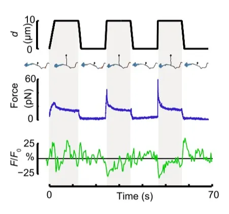
Time dependence of trap position (top), force signal (middle) and Ca2+ fluorescence signal (bottom) recorded during a membrane tether pulling experiment. The force and Ca2+ ion channel activity signals exhibit negative correlation.

Fig. 2

Force and displacement data for two beads stimulated at frequencies of 4 Hz and 0.25 Hz. The progressive reduction of the beads displacement is in concomitancy with an increase of the applied force and loading rate.

Fig. 1

Schematic representation of the optical tweezers experiment. A fibronectin-coated bead is trapped, brought into contact with the cell membrane and stimulated with oscillations at different frequencies.

Fig.2

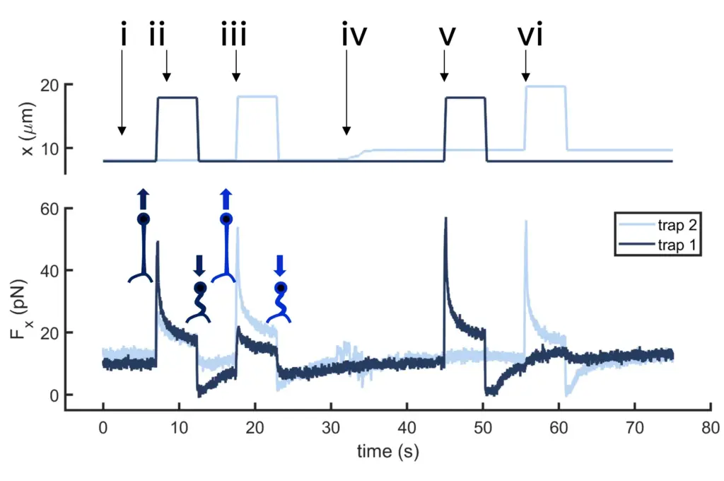
Time dependence of trap 1 and trap 2 position (top) and force (bottom) data along the different steps of the described dual tether pulling experiment.

Fig.1

Confocal video of the described dual tether pulling assay performed on a neuron axon. Courtesy of M. Krieg lab (ICFO).

Fig. 1

Time dependence of trap position (top), force signal (middle) and Ca2+ fluorescence signal (bottom) recorded during a membrane tether pulling experiment. The force and Ca2+ ion channel activity signals exhibit negative correlation.

Video 1

Confocal video showing a DVA neuron Ca2+ ion channel activity in a dynamic optical trapping assay. The pulling rate applied to the membrane tether is progressively increased. Scale bar = 5µm. Acquired at 10Hz.