APPLICATIONS / OPTICAL TWEEZERS

Applications of SENSOCELL +
optical tweezers

SENSOCELL+ combines direct force measurements with fluorescence imaging and optional integrated high-speed confocal microscopy for correlated quantitative assays across molecular, cellular and soft-matter systems. Explore applications in mechanobiology, molecular biophysics, biomolecular condensates and soft matter physics.

01 / RESEARCH FIELDS

From molecular interactions to living mechanics

Explore optical trapping applications across the systems you study, from individual molecules and biomolecular condensates to living cells, tissues and complex soft matter.

01

CELL MECHANOBIOLOGY

Forces, mechanics and interactions in living cells and tissues.

02

MOLECULAR BIOPHYSICS

Motors, filaments and single-molecule interactions.

03

PHASE SEPARATION

Mechanical evolution of biomolecular condensates.

04

SOFT MATTER PHYSICS

Colloids, gels, biofilms and complex fluids.

01

CELL MECHANOBIOLOGY

Forces, mechanics and interactions in cells, tissues and living organisms.

02

MOLECULAR BIOPHYSICS

Molecular interactions, motor proteins and cytoskeletal filaments.

03

BIOMOLECULAR CONDENSATES

Phase separation, viscoelasticity and fusion dynamics.

04

SOFT MATTER PHYSICS

Colloids, gels, biofilms and active materials.

02 / MECHANOBIOLOGY

Live-cell mechanics across scales

Measure membrane tension, intracellular mechanics and rheology, cell-cell interactions and force generation in living cells, tissues and organisms.

MEMBRANE TENSION & RECEPTOR FORCES

Measure membrane tension and receptor forces with automated tether pulling and particle-cell interaction assays.

NUCLEUS & SUBCELLULAR MECHANICS

Apply creep and stress-relaxation assays to probe nuclear and cytoplasmic mechanics inside living cells.

INTRACELLULAR RHEOLOGY

Quantify the viscoelastic properties of the cytoplasm, nucleus and other intracellular environments.

CELL-CELL INTERACTIONS

Measure immune cell binding forces with control over cell orientation and contact time.

CELL STIFFNESS & ELASTICITY

Indent cells to quantify the stiffness, elasticity and viscoelastic response of the cell membrane and cortex.

CELL FORCE DYNAMICS

Measure the forces generated by cells and the mechanical work they produce.

03 / MOLECULAR BIOPHYSICS

Measure forces and dynamics at the molecular scale

Use SENSOCELL+ to quantify molecular interactions, characterize cytoskeletal mechanics and resolve the activity of individual molecular motors through direct force measurements and correlated fluorescence imaging. 

MOTOR PROTEINS & CYTOSKELETAL FILAMENTS

Measure motor protein activity and characterize the mechanics of cytoskeletal filaments and networks in vitro and inside living cells. 

SINGLE-MOLECULE FORCE SPECTROSCOPY

Apply and measure forces in dual-trap and three-bead assays, from DNA stretching to individual myosin motor dynamics. 

04 / BIOMOLECULAR CONDENSATES

Follow condensates
mechanics as they age

Track changes in material state as phase-separated condensates form, fuse and age. 

MECHANICS OF BIOMOLECULAR CONDENSATES

Quantify bulk and interfacial viscoelasticity through droplet fusion analysis and active microrheology. 

05 / SOFT MATTER PHYSICS

Complex materials.
Their mechanics, simplified.

Explore how interactions and viscoelasticity govern the behavior of complex and active soft-matter systems. 

COLLOIDS, GELS, BIOFILMS & ACTIVE MATERIALS

Measure interaction forces and complex shear modulus using automated force-probing and active microrheology workflows. 

06 / YOUR APPLICATION

Is your application
not listed above?

If your application is not listed, discuss it directly with our scientists. We work with research teams to assess feasibility and define workflows.

Fig. 1

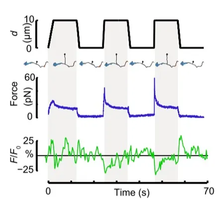
Time dependence of trap position (top), force signal (middle) and Ca2+ fluorescence signal (bottom) recorded during a membrane tether pulling experiment. The force and Ca2+ ion channel activity signals exhibit negative correlation.

Fig. 2

Force and displacement data for two beads stimulated at frequencies of 4 Hz and 0.25 Hz. The progressive reduction of the beads displacement is in concomitancy with an increase of the applied force and loading rate.

Fig. 1

Schematic representation of the optical tweezers experiment. A fibronectin-coated bead is trapped, brought into contact with the cell membrane and stimulated with oscillations at different frequencies.

Fig.2

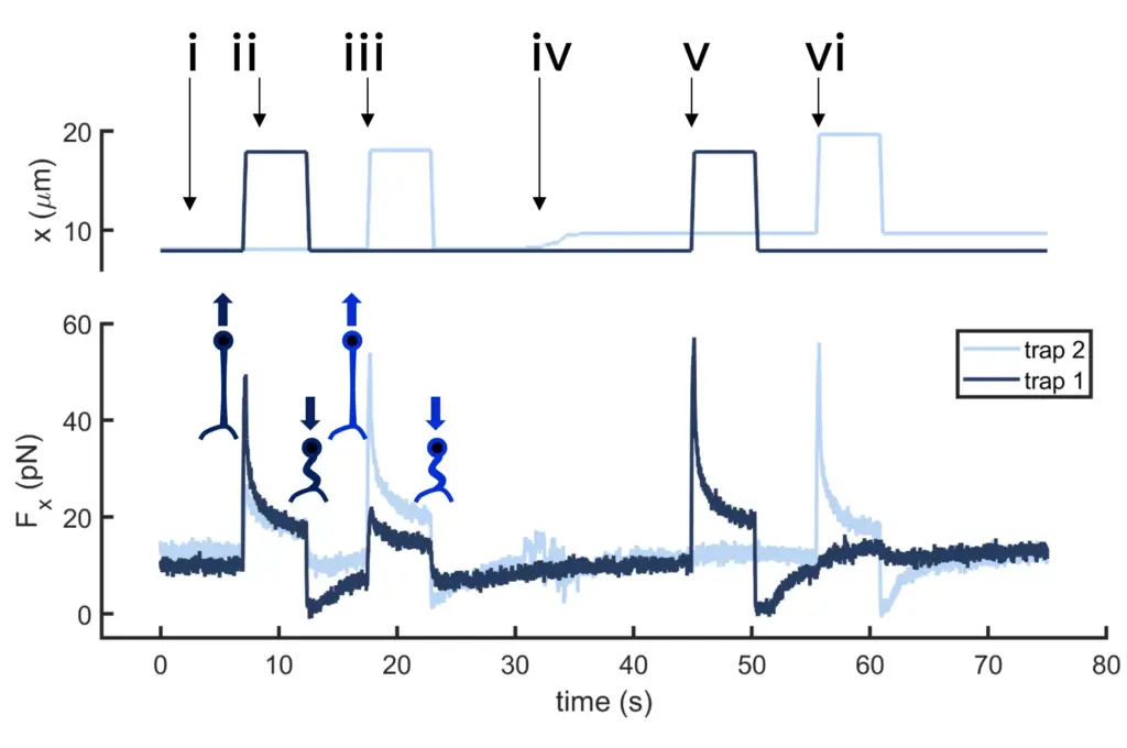
Time dependence of trap 1 and trap 2 position (top) and force (bottom) data along the different steps of the described dual tether pulling experiment.

Fig.1

Confocal video of the described dual tether pulling assay performed on a neuron axon. Courtesy of M. Krieg lab (ICFO).

Fig. 1

Time dependence of trap position (top), force signal (middle) and Ca2+ fluorescence signal (bottom) recorded during a membrane tether pulling experiment. The force and Ca2+ ion channel activity signals exhibit negative correlation.

Video 1

Confocal video showing a DVA neuron Ca2+ ion channel activity in a dynamic optical trapping assay. The pulling rate applied to the membrane tether is progressively increased. Scale bar = 5µm. Acquired at 10Hz.