BLOG

Extending calibration-free force measurements to optically-trapped rod-shaped samples

Extending calibration-free force measurements to optically-trapped rod-shaped samples

An interesting new article have been published today in Scientific Reports related with IMPETUX technology:

Extending calibration-free force measurements to optically-trapped rod-shaped samples – Català, F. et al.

In this article the authors demonstrate that the beam momentum detection can be applied to measure forces on optically trapped cylinders, without the need for previous trap calibration or complete understanding of the trapping dynamics.

Since Force measurement based on beam momentum detection is independent of any local parameter present in the experiment, in addition to rod-shaped samples, the method described and based on IMPETUX patented Technology can be undertaken in non-viscous media, such an interior of a Cell. Biological cargos driven by molecular motors can be directly trapped without the aid of spherical probes, and the pulling forces measured under strict physiological conditions.

For more detailed information , have a look at the article here.

Enjoy the reading!
More news:

Fig. 1

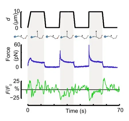
Time dependence of trap position (top), force signal (middle) and Ca2+ fluorescence signal (bottom) recorded during a membrane tether pulling experiment. The force and Ca2+ ion channel activity signals exhibit negative correlation.

Fig. 2

Force and displacement data for two beads stimulated at frequencies of 4 Hz and 0.25 Hz. The progressive reduction of the beads displacement is in concomitancy with an increase of the applied force and loading rate.

Fig. 1

Schematic representation of the optical tweezers experiment. A fibronectin-coated bead is trapped, brought into contact with the cell membrane and stimulated with oscillations at different frequencies.

Fig.2

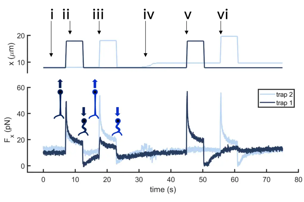
Time dependence of trap 1 and trap 2 position (top) and force (bottom) data along the different steps of the described dual tether pulling experiment.

Fig.1

Confocal video of the described dual tether pulling assay performed on a neuron axon. Courtesy of M. Krieg lab (ICFO).

Fig. 1

Time dependence of trap position (top), force signal (middle) and Ca2+ fluorescence signal (bottom) recorded during a membrane tether pulling experiment. The force and Ca2+ ion channel activity signals exhibit negative correlation.

Video 1

Confocal video showing a DVA neuron Ca2+ ion channel activity in a dynamic optical trapping assay. The pulling rate applied to the membrane tether is progressively increased. Scale bar = 5µm. Acquired at 10Hz.