
Study the role of cell nucleus mechanotransduction pathways and how the nucleus mechanical properties regulate cellular activity such as cell migration, development or disease.
Characterize the mechanical properties of intracellular medium like the cell nucleus or the cytoplasm and obtain information of its rigidity, viscosity and elasticity for different cell types and conditions. Investigate how diseases like cancer or laminopathies affect nuclear mechanics and how these influence and reflect cellular differentiation and development.
Use Sensocell optical tweezers to perform cell nucleus indentation experiments inside living cells, embryos or organisms using internalized microspheres or subcellular structures such as lipid droplets as probes. Stress-relaxation tests involve applying a constant deformation to the nucleus and observing how the force (or stress) relaxes over time.
The bead or the lipid droplet is trapped and used to indent the nucleus to a certain depth and held at that position.
Over time, the force required to maintain the indentation decreases as the nucleus relaxes. This force relaxation is recorded.
1. Viscoelastic properties: the relaxation behavior provides insights into the viscoelastic properties of the nucleus. A common approach is to fit the relaxation data to a viscoelastic model, such as the Maxwell or Kelvin-Voigt models.
2. Time constants: these models provide time constants that describe how quickly the nucleus relaxes, which is related to its viscosity and elasticity.
Creep tests involve applying a constant force to the probed material and observing how the deformation (strain) evolves over time. These tests are particularly useful for understanding the long-term viscoelastic behavior of the nucleus or the cell cytoplasmic medium.
V. Venturi et al.
The nucleus measures shape changes for cellular proprioception to control dynamic cell behavior.

Frederic Català-Castro et al.
Direct Force Measurements of Subcellular Mechanics in Confinement using Optical Tweezers.

Frederic Català-Castro, Santiago Ortiz-Vásquez, Carmen Martínez-Fernández et al.
Active microrheology with a single, time-shared laser trap.De Lope-Planelles, A., González-Novo, R., Madrazo, E. et al.

Mechanical stress confers nuclear and functional changes in derived leukemia cells from persistent confined migration.The following images and videos are courtesy of the lab of Dr. Verena Ruprecht (CRG) in collaboration with Dr. Stefan Wieser and Dr. Michael Krieg (ICFO). This example is part of a work (Science, 2020) dealing with the role of nucleus mechanotransduction pathways to control cell deformation behavior and cell migration plasticity. The authors used SENSOCELL optical tweezers to optically trap and manipulate 1 μm fluorescent beads and indent the nuclei of stem cells extracted from zebrafish embryos. The beads had been previously internalized into the 1-cell early embryos. For the experiments, the authors used automatized Stress-Relaxation routines consisting in predefined back and forth trap motions.
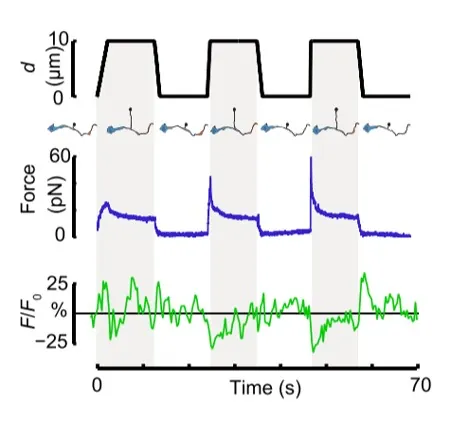
Before launching the Stress-Relaxation routine, the optically trapped bead is some microns away from the nucleus membrane. When we launch the routine, the optical trap moves towards the nucleus at a certain speed until it reaches the nucleus membrane and the indentation takes place. At this point, the optical trapping force increases from zero and gives rise to a force peak. The indentation depth depends on the predefined optical trap trajectory. In this case, the routine was programmed in such a way that the optical trap remained indenting the cell nucleus for 10s. During these 10 seconds, we observe a relaxation in the force signal. Next, the optical trap goes back to its original position and the recorded force drops down to zero. Figure 2 shows the optical trap time-position and time-force curves for a single cell nucleus indentation experiment.
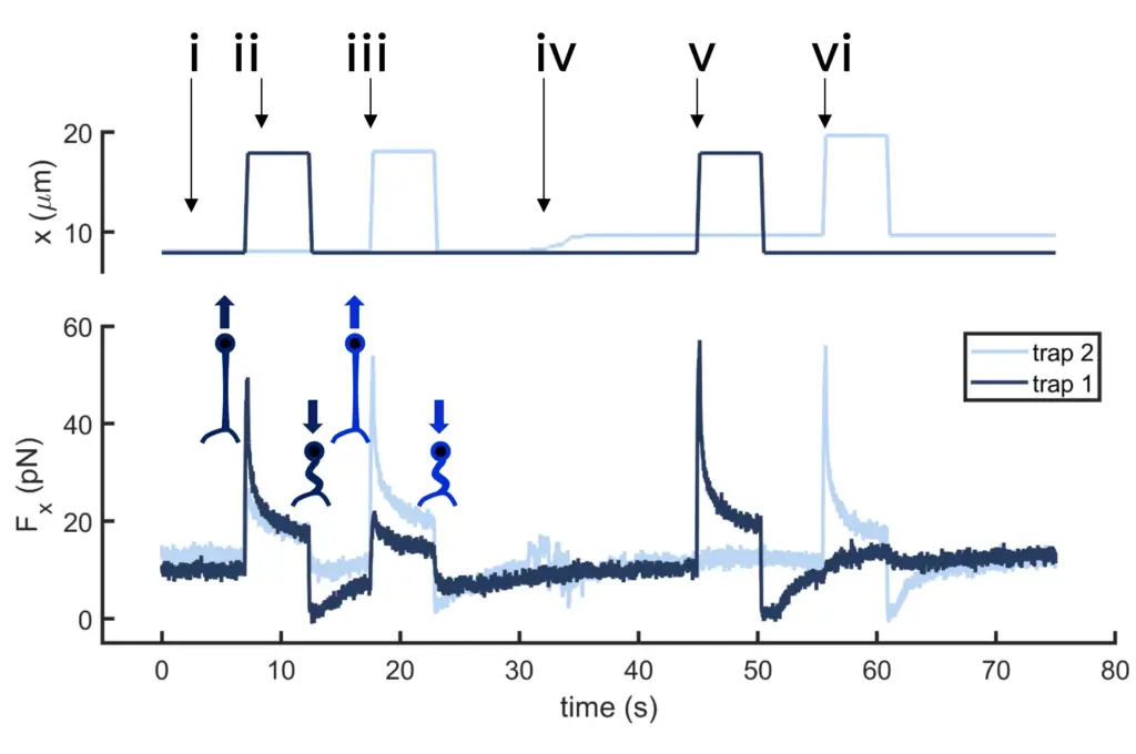
If the tethers are close enough, data reveals tension propagation across the membrane, seen in the force peak in trap 2 when only the first tether is pulled and vice versa. However, when the tethers are far apart, this phenomenon disappears. Thus, tuning the tether-to-tether distance quantifies how cell membrane tension decays with distance.
Below, we can see some results obtained from an exponential-decay fitting of such force relaxation curves for cells in suspension and cells confined in microchambers. From these fittings the authors obtain the characteristic relaxation time and the static force given by the residual stress applied onto the nuclear membrane for both conditions.
This is an example of a cell nucleus indentation experiment performed inside the intestinal cells of a living C. elegans organism (Biorxive 2023). Courtesy of Michael Krieg’s lab (ICFO). In this work the researchers show how endogenous lipid droplets are excellent probes to perform intracellular mechanic experiments with Sensocell optical tweezers, including nucleus deformation by stress-relaxation assays.
The following images and data illustrate a dual-trap cell nucleus indentation experiment conducted using a pair of 1 μm beads internalized in zebrafish stem cells. Each bead indents the cell nucleus sequentially at different times. The system continuously records the force and position data of both traps, enabling the detection of any force transmission between them. While control cells do not exhibit force transmission during nucleus indentation, cells treated with Latrunculin A for actin depolymerization show a distinct force propagation signature. This indicates that actin plays a crucial role in stabilizing the position of the cell nucleus within the cell.
Here, the authors present a protocol to investigate the intracellular mechanical properties of isolated embryonic zebrafish cells in three-dimensional confinement with direct force measurement by an optical trap.
Introduction / Single-cell Preparation and Staining / Optical Trapping (OT) Chamber Spacing and Optical Tweezers Start-up / Alignment of the Optical Force Sensor / Performing the Nucleus Indentation Experiments / Results: Measurement of the Forces and Material Properties of the Cell Nucleus Inside Zebrafish Embryo / Conclusion. See full video here.
In this video example, the vacuole of a yeast cell was optically trapped and used to probe the cellular medium in two ways:
First, we perform a Creep test: the force feedback routine (force clamp) tracks the cell’s vacuole movement while a constant force of 5 pN is applied on it. The data from the plot panel show force and position signals (orange and blue lines respectively). When the force clamp is applied, the position signal follows the classical behaviour expected for a viscoelastic medium. When the force clamp is stopped, the force signal relaxes back to zero (negative values) and the position signal abruptly shifts to zero.
Next, we launch a Stress-Relaxation test using the same trapped vacuole as a probe. In this case, we apply a trajectory to the optical trap to move it 300 nm to the right in 0.1 s while the applied force is tracked. We can observe a peak in the force when the trap position is shifted followed by a relaxation of the force while the trap remains in a fixed location.
In summary, Sensocell optical tweezers is a powerful tool for measuring the mechanical properties of the cell nucleus. Indentation experiments provide data on stiffness and elasticity, while stress-relaxation tests reveal viscoelastic properties, offering comprehensive insights into nuclear mechanics.
Time dependence of trap position (top), force signal (middle) and Ca2+ fluorescence signal (bottom) recorded during a membrane tether pulling experiment. The force and Ca2+ ion channel activity signals exhibit negative correlation.
Force and displacement data for two beads stimulated at frequencies of 4 Hz and 0.25 Hz. The progressive reduction of the beads displacement is in concomitancy with an increase of the applied force and loading rate.
Schematic representation of the optical tweezers experiment. A fibronectin-coated bead is trapped, brought into contact with the cell membrane and stimulated with oscillations at different frequencies.
Time dependence of trap 1 and trap 2 position (top) and force (bottom) data along the different steps of the described dual tether pulling experiment.
Confocal video of the described dual tether pulling assay performed on a neuron axon. Courtesy of M. Krieg lab (ICFO).
Confocal video showing a DVA neuron Ca2+ ion channel activity in a dynamic optical trapping assay. The pulling rate applied to the membrane tether is progressively increased. Scale bar = 5µm. Acquired at 10Hz.