
Explore the dynamics of soft matter and colloid properties of emulsions, gels, biomaterials and liquid crystals with SENSOCELL optical tweezers.
Optical tweezers can measure the force-displacement relationship of a colloidal particle in a fluid or gel, by applying a controlled displacement to the trapped particle and measuring the resultant force. By monitoring the particle’s response to these forces (e.g., its position fluctuations), researchers can deduce the viscoelastic G modulus (elastic and viscous components) of the surrounding medium.
Additionally, Sensocell optical tweezers allow for microrheological experiments by tracking the movement of colloidal particles and assessing how the medium resists or responds to deformation. This technique is particularly valuable in complex fluids like biological gels or polymers, where traditional rheology methods are difficult to apply.

Hunter Seyforth, Mauricio Gomez, W. Benjamin Rogers, Jennifer L. Ross, and Wylie W. Ahmed.
Nonequilibrium fluctuations and nonlinear response of an active bath.
Ophelie Squillace, Ting Zhang, Lia Argentou, Andrew Ward, Zhenyu J. Zhang.
Nanoscopic wetting behaviour of single oil droplets on a fibre.In this example, Dr Ahmed (Université de Toulouse; Slam-Lab) et al., taken from the paper titled “Nonequilibrium fluctuations and nonlinear response of an active bath“ explore the dynamics of a passive colloidal probe immersed in an active bath composed of E. coli bacteria.
The authors use optical tweezers and light momentum analysis to study the interactions between the colloidal probe and the surrounding active bath, comparing these results with those obtained from a passive bath (water).
This research provided experimental evidence of how active matter, such as a suspension of E. coli, can induce complex, nonequilibrium dynamics in immersed particles. This has implications for understanding the behavior of systems far from equilibrium.
The passive probe in the active bath exhibited enhanced nonequilibrium fluctuations compared to the passive bath. These fluctuations are driven by the activity of the bacteria and deviate significantly from what is expected in equilibrium systems. The authors measured the active energy spectrum for the quantification of the nonthermal energetic fluctuations of a colloid in the active bath
The viscosity of the active bath varied with the Péclet number (Pe), a dimensionless number representing the ratio of advective to diffusive transport rates. The study observed shear thinning at low Pe values, followed by shear thickening at intermediate Pe values, and a plateau in viscosity at high Pe values. This behavior contrasts with the passive bath, where no such variations were noted.
The distribution of force fluctuations in the active bath depended on Pe, with higher Pe leading to wider distributions, indicating more significant deviations from thermal equilibrium.
This example taken from the paper titled “Nanoscopic wetting behaviour of single oil droplets on a fibre” published in Colloids and Surfaces A Journal. In this work, researchers from from the University of Birmingham and the Central Laser Facility at the Rutherford Appleton Laboratory studied the interactions between silicone oil droplets and microscopic fibers, focusing on the wetting and surface interactions under various conditions. Using optical tweezers combined with our direct force sensor based on momentum analysis, the researchers quantified these interactions.
The study examined how factors such as surfactant chemistry, pH, and salt concentration influence the behavior of silicone droplets when they come into contact with different fiber types, particularly cotton and polyester.
The interaction between the oil droplets and fibers is largely influenced by the electrostatic forces, which can be modulated by changing the salt concentration in the surrounding medium. Indeed, higher salt concentrations reduced the electrostatic repulsion, leading to increased adhesion and spreading of droplets on the fibers. Different fibers exhibited varying degrees of interaction, with polyester showing stronger negative surface charge compared to cotton, affecting how the droplets interacted with each fiber type.
The study successfully demonstrated that optical tweezers could be used to directly measure and quantify these interactions at a microscopic scale, providing valuable insights into the wetting behavior of emulsion droplet colloids on fibers.
Images are courtesy of Andy Ward (CLF Lasers for Science Facility, UK).
In this experiment, 3-micron latex beads were added to different water:glycerol mixtures (0%, 20%, 40%, 60% and 80% glycerol in water) and used as probes to characterize the rheological properties of the gels. This work is in collaboration with BIOPT lab from the University of Barcelona.
For each single experiment, a bead was trapped using our SENSOCELL optical tweezers and forced to oscillate sinusoidally with 200 nm amplitude at increasing frequencies (3Hz to 100 Hz) launching our TimSom acive rheology built-in routine. The calculated viscosity values were then obtained by fitting the frequency dependence of the measured loss modulus (G”).
In a similar fashion, we performed active micro-rheology experiments inside polyacrylamide gels with embedded micrometer beads to get their complex shear moduli.
Sensocell is a powerful tool in the field of colloid physics, particularly for studying the mechanical and dynamic properties of colloidal particles. Its ability to manipulate particles with high precision and minimal disturbance, combined with the potential for real-time and quantitative analysis, allows for a deeper understanding of colloidal systems.
Confocal video of the described dual tether pulling assay performed on a neuron axon. Courtesy of M. Krieg lab (ICFO).
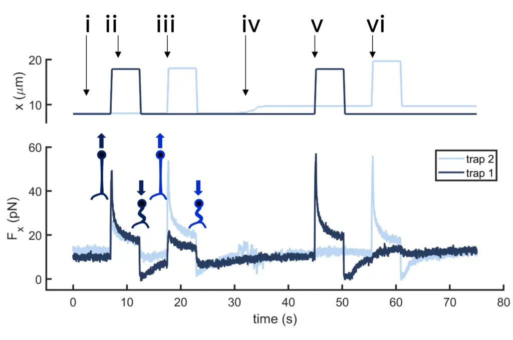
Time dependence of trap 1 and trap 2 position (top) and force (bottom) data along the different steps of the described dual tether pulling experiment.
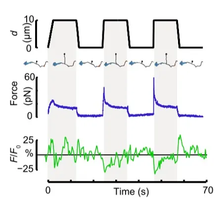
Time dependence of trap position (top), force signal (middle) and Ca2+ fluorescence signal (bottom) recorded during a membrane tether pulling experiment. The force and Ca2+ ion channel activity signals exhibit negative correlation.
Force and displacement data for two beads stimulated at frequencies of 4 Hz and 0.25 Hz. The progressive reduction of the beads displacement is in concomitancy with an increase of the applied force and loading rate.
Schematic representation of the optical tweezers experiment. A fibronectin-coated bead is trapped, brought into contact with the cell membrane and stimulated with oscillations at different frequencies.
Time dependence of trap position (top), force signal (middle) and Ca2+ fluorescence signal (bottom) recorded during a membrane tether pulling experiment. The force and Ca2+ ion channel activity signals exhibit negative correlation.
Confocal video showing a DVA neuron Ca2+ ion channel activity in a dynamic optical trapping assay. The pulling rate applied to the membrane tether is progressively increased. Scale bar = 5µm. Acquired at 10Hz.