Optical tweezers are a powerful tool for studying liquid-liquid phase separation (LLPS) in biomolecular condensates and quantifying their mechanical properties.
Optical tweezers can track changes in the mechanical properties of protein condensates over time, revealing their aging and maturation, transitioning from liquid-like to solid-like states.
This method uses two trapped beads to sandwich
the condensate, allowing for detailed measurements
of surface tension and shear modulus at the
protein droplet interface region.
This patented method integrated in SENSOCELL’s workflow, uses a single bead embedded within the protein droplet and a single optical trap to apply forces and measure responses, thus simplifying the experimental setup. The TimSOM active rheology routine automatically computes the complex G modulus (G=G’+iG”) of the material. Sensocell allows measuring the G modulus over 5 orders of magnitude of frequency spectrum.
Rheology assays help understanding how pathological conditions affect the mechanical properties of condensates, potentially leading to diseases like neurodegenerative disorders where protein aggregation and stiffening are common. Additionally, testing potential therapeutic compounds’ effects on the mechanical properties of biomolecular condensates is key for developing treatments that can modulate phase transitions and mitigate diseases related to LLPS phenomena.
Sanfeliu-Cerdán, N., Català-Castro, F., Mateos, B. et al.
A MEC-2/stomatin condensate liquid-to-solid phase transition controls neuronal mechanotransduction during touch sensing.
Frederic Català-Castro, Santiago Ortiz-Vásquez, Carmen Martínez-Fernández et al.
Active microrheology with a single, time-shared laser trap.This study led by Michael Krieg’s lab (ICFO) explores the use of TimSOM Rheology integrated in SENSOCELL optical tweezers to measure the frequency-dependent mechanical properties of viscoelastic protein droplets undergoing LLPS. This process is critical for organizing proteins within the cytoplasm of living cells and is tightly regulated to ensure proper cellular functions.
Uncontrolled transitions of these condensates into stiffened states are often linked to diseases. Indeed, increased stiffness correlates with disease severity. The researchers applied TimSOM rheology on MEC-2 condensates, comparing it to the dual trap assay.
The findings show that both configurations detected a shift in the crossover frequency to lower values after 24 hours of protein droplet formation, indicating maturation. However, the single trap assay (TimSOM) recorded slightly higher stiffness and viscosity, suggesting non-uniform maturation across the droplet. TimSOM effectively resolved the fractional Maxwell behavior of the condensates and revealed a mechanism for rigidity transition across the condensate.
The rheological data of Fig.1 support these findings, showing the storage (G′) and loss (G′′) moduli at different times demonstrating changes in viscosity, stiffness, time constant, and crossover frequency over 24 hours of condensate maturation.
These results confirm that SENSOCELL optical tweezers can accurately measure the viscoelastic properties of aging biomolecular condensates, potentially aiding in drug screening applications.

SENSOCELL optical tweezers are an invaluable tool for researchers studying liquid-liquid phase transitions in biomolecular condensates. They provide high-resolution, quantitative data on the viscoelastic properties of these structures, helping to unravel the mechanisms underlying their formation, maturation, and role in cellular function and disease.
Confocal video of the described dual tether pulling assay performed on a neuron axon. Courtesy of M. Krieg lab (ICFO).
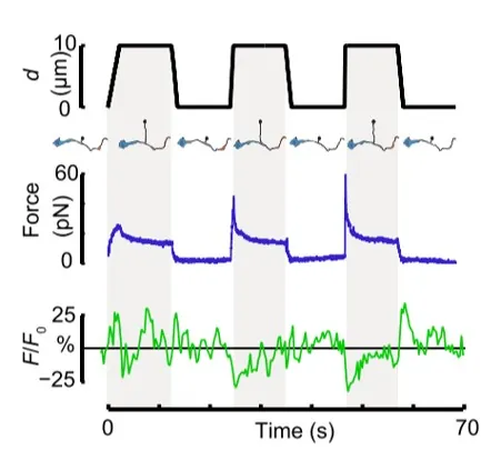
Time dependence of trap position (top), force signal (middle) and Ca2+ fluorescence signal (bottom) recorded during a membrane tether pulling experiment. The force and Ca2+ ion channel activity signals exhibit negative correlation.
Force and displacement data for two beads stimulated at frequencies of 4 Hz and 0.25 Hz. The progressive reduction of the beads displacement is in concomitancy with an increase of the applied force and loading rate.
Schematic representation of the optical tweezers experiment. A fibronectin-coated bead is trapped, brought into contact with the cell membrane and stimulated with oscillations at different frequencies.
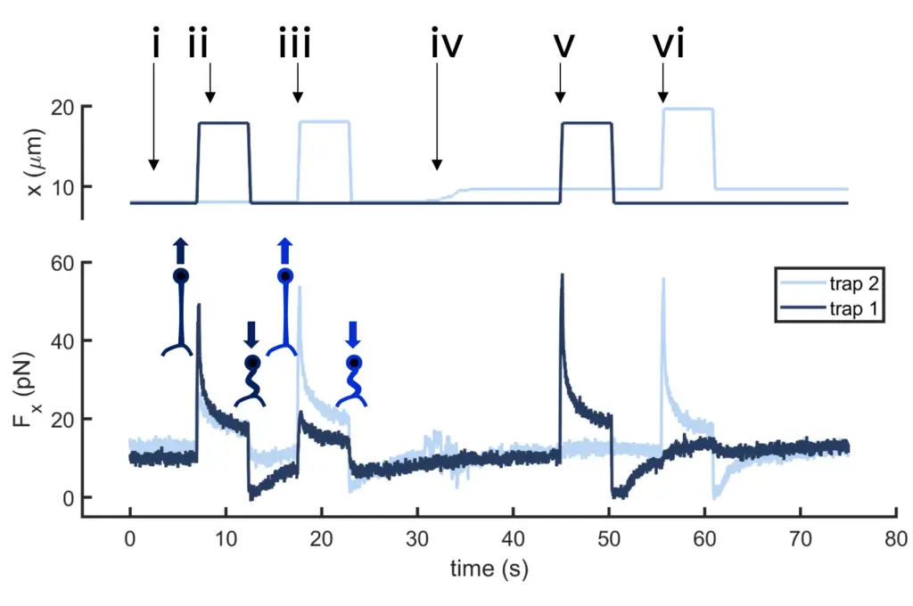
Time dependence of trap 1 and trap 2 position (top) and force (bottom) data along the different steps of the described dual tether pulling experiment.
Time dependence of trap position (top), force signal (middle) and Ca2+ fluorescence signal (bottom) recorded during a membrane tether pulling experiment. The force and Ca2+ ion channel activity signals exhibit negative correlation.
Confocal video showing a DVA neuron Ca2+ ion channel activity in a dynamic optical trapping assay. The pulling rate applied to the membrane tether is progressively increased. Scale bar = 5µm. Acquired at 10Hz.